

A tender disc under the nipple, a soft chest after weight gain, and a puffy areola can look similar in a mirror. They can come from different tissues. One may be proliferating gland. Another may be fat over a small normal gland. A third may reflect loose skin, scar, swelling, or the shape of the pectoral muscle. The distinction determines the next step.
Gynecomastia is benign growth of male breast glandular tissue. Many causes can produce it. Pseudogynecomastia describes breast enlargement dominated by fat without the characteristic glandular proliferation. Mixed presentations are common. Current European and Italian guidelines begin with history and physical examination because a hormone value cannot establish which of these structures is present.[S1][S2]
The tissue also has a history. Human pathology shows heterogeneous ductal and stromal patterns. Some specimens appear more proliferative. Others contain denser fibrosis. These patterns help explain why tissue that began or enlarged within six months can behave differently from adult tissue present beyond six to twelve months. A calendar still cannot identify an individual’s histology or guarantee response.[S3]
Blood tests remain useful. They can reveal hypogonadism, hyperthyroidism, hyperprolactinemia, an hCG signal, liver or kidney disease, and other contexts that change management. They also have clear limits. Serum testosterone, estradiol, prolactin, DHT, or a testosterone-to-estradiol ratio does not measure local enzyme activity, receptor abundance, receptor function, or the tissue’s accumulated response. A normal result today cannot erase an earlier exposure.
Treatment can aim to prevent new growth, relieve pain, stop progression, shrink the gland, or correct the chest contour. Studies need to state which result they measured because less tenderness says little about how much gland remains. Lower estradiol alone does not show that the gland became smaller. Surgery can improve the contour while an endocrine cause still needs care.
Diagnosis also starts with the presentation. A typical, symmetric, central presentation often needs no imaging. An indeterminate mass or a suspicious presentation follows a different route based on age and examination. Hard or eccentric tissue, visible or measurable unilateral enlargement over days to weeks, nipple retraction or spontaneous discharge, skin ulceration or dimpling, enlarged axillary nodes, and a testicular abnormality leave the observation-and-drug pathway and enter diagnostic assessment. No validated centimeter-per-week cutoff separates benign from malignant growth. The American College of Radiology makes this presentation-based distinction explicit.[S4]
Trials are only one layer of the treatment record. Patents reveal local-delivery ideas and proposed combinations. Forums reveal nonresponse, adverse effects, mechanism switches, and the practical preference for raloxifene in bodybuilding. Human studies still control claims about efficacy. The other layers now shape the protocol and show where its certainty changes.
The time labels are fixed throughout this guide. Recent adult disease means documented onset or enlargement within six months. Structurally established adult disease means tissue present beyond six to twelve months. Persistent after cause correction means tissue still present after at least six months of observation following correction of the identified cause. Rapid enlargement means visible or measurable change over days to weeks. Early postoperative means the first six weeks. These are decision anchors, not histology tests.
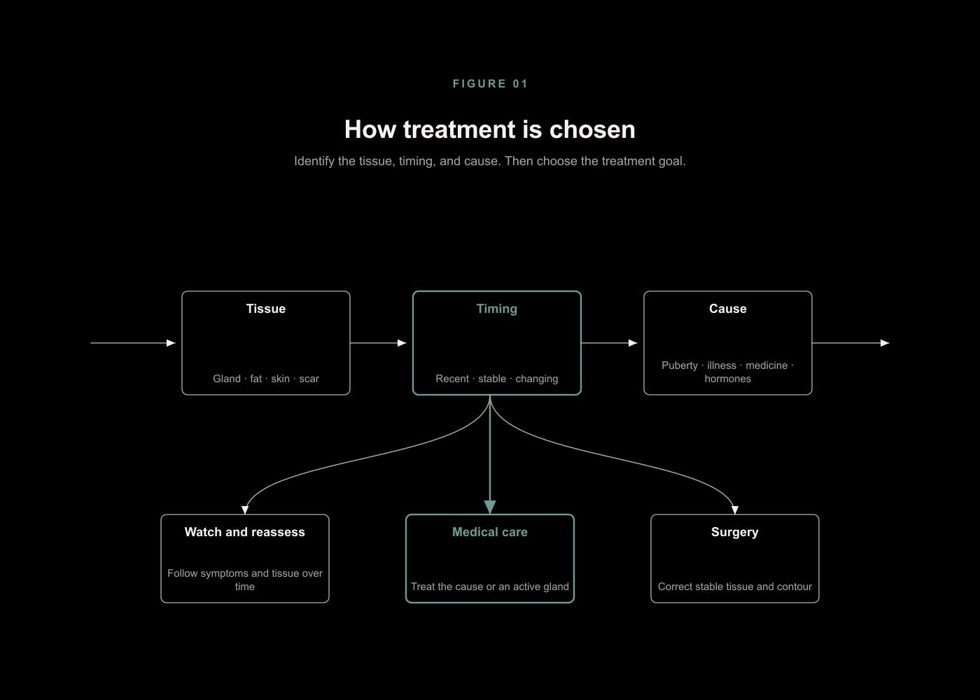
Figure 1. Treatment is chosen from the tissue, timing, cause, and the result the patient needs.[S1][S2][S3][S6]
Contents
Part I. Identify the tissue
1. Male breast anatomy.
2. Gland, fat, skin, scar, and muscle.
3. Tissue change over time.
4. Clinical grade and structural anatomy.
Part II. Explain the biology
5. Estrogenic stimulation and androgenic opposition.
6. Serum values and breast-tissue exposure.
7. Local steroid metabolism.
8. DHT and other 5-alpha-reduced steroids.
9. Prolactin, progesterone, and progestins.
10. Growth factors, inflammation, and fibrosis.
Part III. Find the cause
11. Physiological life stages.
12. Obesity, weight change, nutrition, and recovery.
13. Hypogonadism and genetic conditions.
14. Thyroid, liver, kidney, and systemic disease.
15. Tumors and urgent endocrine causes.
16. Medication-induced gynecomastia.
17. TRT, AAS, fertility treatment, and changing exposure.
18. Idiopathic disease after complete evaluation.
Part IV. Diagnose and classify
19. Urgent features and malignant differentials.
20. The exposure and symptom timeline.
21. Physical examination.
22. Targeted laboratory evaluation.
23. Imaging and biopsy.
24. Final diagnostic classification.
Part V. Choose the treatment objective
25. Prevention, pain relief, growth arrest, regression, and removal.
26. Observation and cause correction.
27. Objective reassessment.
Part VI. Medical treatment
28. SERMs.
29. Aromatase inhibitors.
30. DHT and historical androgen treatment.
31. Danazol, mesterolone, and older strategies.
32. Dopamine agonists.
33. Cause-specific medical paths.
34. Treatment safety and stopping rules.
Part VII. Supplements and nonstandard treatment
35. Supplements linked to estrogen metabolism.
36. Diet and metabolic claims.
37. Topical and physical approaches.
38. Patent claims.
39. Forum, Reddit, and social evidence.
Part VIII. Surgery
40. Surgical indications.
41. Anatomy-selected operations.
42. Adequate excision and contour preservation.
43. Evaluating a surgeon and operative plan.
44. Complications and recovery.
45. Limits of the surgical outcome literature.
Part IX. Residual tissue, recurrence, and treatment failure
46. Residual gland, fat, scar, skin, and contour.
47. Medical nonresponse and changing diagnosis.
48. Early postoperative swelling and scar maturation.
49. New gland growth after renewed exposure.
50. Revision evaluation and long-term follow-up.
Part X. Gradual intervention protocols and common paths
Protocol 0. Establish a usable baseline.
Protocol 1. Remove the active cause and observe.
Protocol 2. Treat a recent active gland with one SERM.
Protocol 3. Consider DHT as a separate mechanism.
Protocol 4. Switch mechanisms after DHT or SERM failure.
Protocol 5. TRT, AAS, hCG, and bodybuilding exposure.
Protocol 6. Historical reserve treatments.
Protocol 7. Stop endocrine trials when the problem is structural.
51. Adolescents.
52. Adults with a recent tender lump.
53. Long-standing stable disease.
54. Obesity and major weight change.
55. TRT and AAS exposure.
56. Fertility treatment.
57. Medication-associated disease.
58. Prostate-cancer antiandrogen treatment.
59. Confirmed endocrine or systemic disease.
60. Postoperative concern about recurrence.
Part XI. Evidence limits and reference tools
61. Evidence hierarchy.
62. Material disagreements.
63. Unresolved questions.
Conclusion. Treatment follows tissue, timing, cause, and goal
Appendix A. Urgent-feature checklist
Appendix B. Exposure and symptom timeline template
Appendix C. Laboratory interpretation framework
Appendix D. Medication-causality worksheet
Appendix E. Treatment-evidence summary
Appendix F. Surgery consultation checklist
Appendix G. Postoperative finding and recurrence taxonomy
Appendix H. Glossary